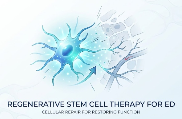
How Stem Cell Therapy Restores Vascular Health in Erectile Dysfunction
Erectile dysfunction is primarily a vascular and neurogenic disorder, even though it is often discussed solely in terms of sexual performance. An erection relies on a precise biological cascade. When sexual arousal occurs, parasympathetic nerve impulses trigger the release of nitric oxide. This biochemical signal relaxes the smooth muscle lining the corpus cavernosum, allowing arterial blood to engorge the sponge-like erectile chambers while veins constrict to trap the blood. When microvascular damage, endothelial breakdown, or nerve injury occurs, the tissue loses its capacity to fill and maintain pressure.
Identifying the primary causes of Erectile dysfunction is critical for determining appropriate long-term interventions. Common contributors include type 2 diabetes, hypertension, atherosclerosis, dyslipidemia, metabolic syndrome, and direct surgical trauma following radical prostatectomy. Over time, chronic vascular impairment starves erectile tissue of oxygen. This persistent lack of perfusion leads to smooth muscle apoptosis (cellular death) and corporal fibrosis, a state where rigid collagen replaces healthy, elastic muscle tissue.
Standard medical treatments do not repair this underlying cellular damage. Phosphodiesterase type 5 (PDE5) inhibitors, such as sildenafil and tadalafil, temporarily block the enzyme that breaks down cyclic guanosine monophosphate (cGMP). While they can enhance blood flow in mild to moderate cases, they depend entirely on having intact vascular tissue to work with, and they do not reverse cellular degeneration. Other interventions, including vacuum constriction devices, intracavernosal alprostadil injections, and surgically implanted prostheses, provide mechanical assistance without restoring natural function. Regenerative medicine focuses on altering this trajectory by targeting tissue repair at the cellular level.
Sourcing and Harvesting Stem Cells: Adipose, Bone Marrow, and Perinatal Tissues
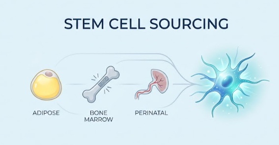
Mesenchymal stem cells (MSCs) are the central biological tools used in regenerative protocols for erectile dysfunction. These cells can be derived from several distinct tissues, each presenting distinct clinical characteristics regarding harvest methods, cell yields, and biological potency.
Autologous Sources: Adipose and Bone Marrow
Autologous stem cells come directly from the patient’s own body, eliminating any risk of donor mismatch or immune rejection.
-
Adipose-Derived Stem Cells (ADSCs): Adipose tissue is harvested via a localized mini-liposuction procedure, typically from the lower abdomen or flanks. Fat tissue contains a high concentration of MSCs within its stromal vascular fraction (SVF), yielding significantly more viable stem cells per gram of tissue than bone marrow aspirate.
-
Bone Marrow-Derived MSCs (BM-MSCs): Bone marrow is obtained through a needle aspiration from the posterior iliac crest of the pelvis. While bone marrow is a historically validated source in orthopedic and hematologic medicine, the extraction procedure is invasive and uncomfortable. Additionally, the proportion of stem cells within fresh bone marrow aspirate is relatively low.
A significant limitation of autologous stem cells is biological senescence. As a person ages, their stem cells experience reduced replication rates, telomere shortening, and diminished cytokine output. In patients with long-standing metabolic disease, hypertension, or advanced vascular disease, autologous cells frequently share the same underlying biological exhaustion that contributed to the erectile dysfunction initially.
Allogeneic Perinatal Tissues: Umbilical Cord Wharton’s Jelly
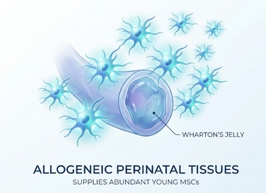
Allogeneic therapies use tissue collected from pre-screened, healthy, full-term donor births. Umbilical cord tissue, particularly the gelatinous substance known as Wharton’s Jelly, provides an abundant supply of young MSCs.
Perinatal stem cells possess youthful cellular characteristics: longer telomeres, faster doubling times, and robust paracrine signaling capacity. Because they do not express mature major histocompatibility complex class II (MHC-II) markers, they are considered immune-privileged, which prevents an adverse immune response in the recipient. Furthermore, allogeneic options eliminate the need for a secondary surgical harvesting procedure, sparing the patient discomfort and recovery time.
Mechanisms of Action in Corpus Cavernosum Repair
Stem cell therapy does not function by transforming injected cells directly into an entirely new organ. Instead, its primary mechanism is paracrine signaling, a process where stem cells release growth factors, cytokines, and extracellular vesicles (exosomes) that instruct local host tissues to initiate repair.
Angiogenesis and Revascularization
Healthy erectile function requires an intact capillary and arterial system. MSCs secrete high levels of pro-angiogenic factors, including vascular endothelial growth factor (VEGF), basic fibroblast growth factor (bFGF), and hepatocyte growth factor (HGF). These signaling molecules stimulate the growth of new capillary networks and repair damaged endothelial cell linings in the helicine arteries, directly restoring arterial inflow into the corpus cavernosum.
Smooth Muscle Regeneration and Anti-Fibrotic Activity
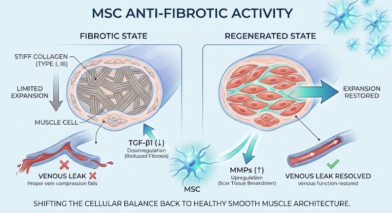
Chronic lack of blood supply leads to the progressive loss of smooth muscle cells, which are gradually replaced by stiff type I and type III collagen. When the corporal body becomes fibrotic, it can no longer expand adequately to compress the subtunical veins, resulting in venous leak. MSCs downregulate transforming growth factor-beta 1 (TGF-beta 1), the primary cytokine responsible for tissue fibrosis, while simultaneously upregulating matrix metalloproteinases (MMPs) to break down excess scar tissue. This shifts the cellular balance back toward healthy smooth muscle architecture.
Cavernous Nerve Regeneration
For men whose erectile dysfunction is neurogenic, such as those recovering from pelvic surgery or suffering from diabetic peripheral neuropathy, MSCs release essential neurotrophic factors. Molecules such as nerve growth factor (NGF) and brain-derived neurotrophic factor (BDNF) encourage axonal sprouting, support Schwann cell survival, and accelerate the repair of damaged cavernous nerves.
Clinical Protocols, Safety, and Synergistic Therapies
Administering stem cell therapy for erectile dysfunction is a focused, outpatient medical procedure designed to maximize local tissue absorption.
Delivery Protocols and Safety
The standard application method involves direct intracavernosal micro-injections. Following the application of a local topical anesthetic or a localized penile nerve block, a clinician uses a fine-gauge needle to distribute the cellular suspension evenly into both chambers of the corpus cavernosum. A light constriction ring or gentle pressure is often applied at the base of the penis for several minutes post-injection to keep the cells concentrated in the erectile tissue before entering systemic circulation. The procedure involves minimal discomfort, and patients can typically resume normal non-strenuous routines within 24 to 48 hours.
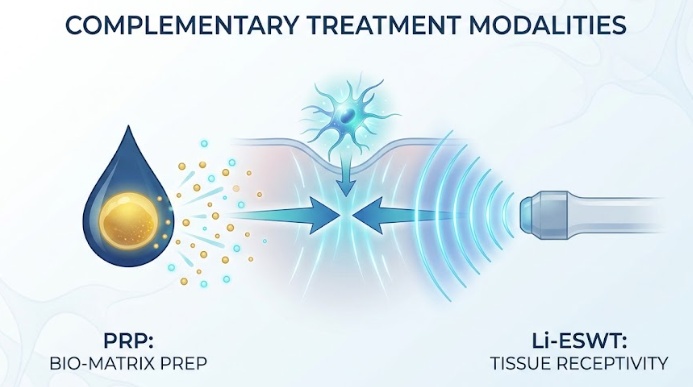
Complementary Treatment Modalities
Clinical protocols frequently combine stem cell injections with supportive therapies to prepare the tissue environment:
-
Platelet-Rich Plasma (PRP): Autologous plasma, concentrated with platelets, provides an immediate release of autologous growth factors that create a supportive bio-matrix for the injected stem cells.
-
Low-Intensity Extracorporeal Shockwave Therapy (Li-ESWT): Acoustic wave therapy delivers focused mechanical pulses to the penile shaft. This induces controlled microtrauma, which upregulates local nitric oxide production, activates local cell receptors, and creates a receptive environment for stem cell integration.
Expected Recovery Timeline
Stem cell therapy requires time to remodel tissue architecture. While individual healing rates vary, structural repair occurs progressively over 6 to 24 weeks. Initial signs of improvement typically begin with the return of involuntary nocturnal and morning erections, followed by improvements in firmness, duration, and response to natural stimulation, as measured by standardized International Index of Erectile Function (IIEF-5) assessments.
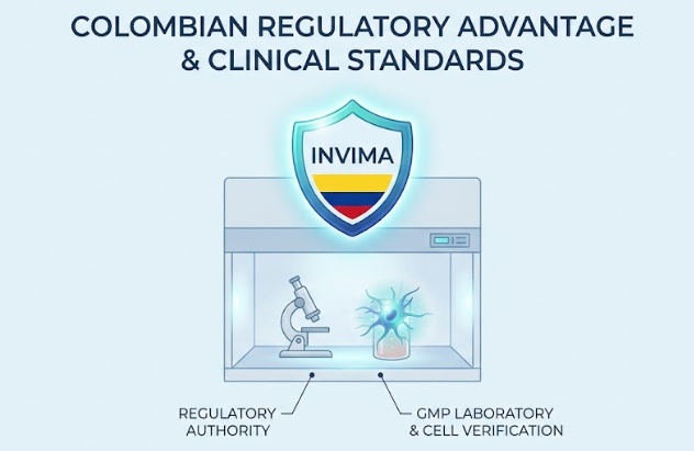
Regulatory Standards and Clinical Advantages in Colombia
Patients seeking advanced regenerative treatments frequently look to Colombia due to its established biotechnology infrastructure and clear regulatory oversight.
In Colombia, cell-based therapies are governed by INVIMA (Instituto Nacional de Vigilancia de Medicamentos y Alimentos), the national regulatory authority overseeing pharmaceuticals and medical procedures. Specialized regenerative clinics operate in coordination with cleanroom facilities certified under Good Manufacturing Practice (GMP) standards. These laboratories carry out strict donor screening, sterility checks, and flow cytometry assays to verify cell count and viability prior to patient administration.
Through adherence to clear regulatory standards and advanced laboratory processing, clinical protocols can utilize high-viability, properly characterized cellular products, providing an evidence-based biological approach to restoring erectile function.