Beyond Blood Flow: The Structural Challenges of the Penis
When men think about erectile health, the conversation usually revolves around blood flow and vascular health. However, a significant number of erectile conditions are strictly structural. While issues like Post-Prostatectomy Erectile Dysfunction often involve nerve damage or altered blood supply, conditions like Peyronie’s disease fall into an entirely different category. They represent structural, non-ischemic pathologies of the penis. Peyronie’s disease involves the development of fibrous scar tissue inside the penis that causes curved, painful erections.
This scar tissue, known as a plaque, forms within a specific anatomical target called the tunica albuginea. The tunica albuginea is the thick, elastic envelope that surrounds the spongy erectile cylinders, known as the corpora cavernosa. Underneath this lies Buck’s fascia, another layer of connective tissue that plays a role in the structural integrity of the penis. When healthy, these tissues expand and stretch smoothly to accommodate the engorgement of blood. When traumatized, they can heal poorly, leading to a cascade of rigid scar formation.
Microvascular Trauma and Plaque Progression
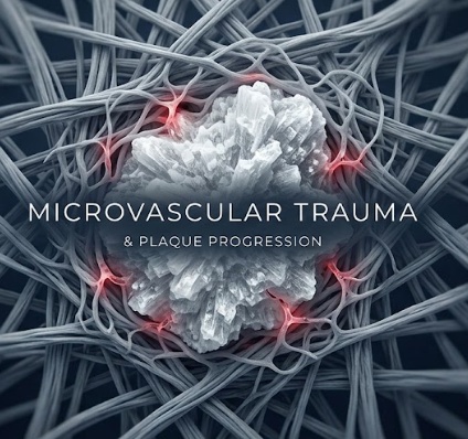
The etiology of Peyronie’s disease almost always points back to microvascular trauma. Repetitive stress during intercourse, sports injuries, or minor accidents can cause microscopic tears in the tunica albuginea. Normally, the body patches these small tears and moves on. However, in men who develop Peyronie’s disease, the healing process becomes severely dysregulated.
The condition typically progresses through two distinct stages. The acute phase involves active inflammation, where patients often experience localized pain regardless of whether they have an erection. Over the course of twelve to eighteen months, the inflammation subsides, leading into the chronic phase. At this point, a stable, calcified plaque has formed, and the severe penile curvature becomes permanent without intervention.
Recognizing the Symptom Profile
The symptoms of penile fibrosis go far beyond a slight bend. Men dealing with this condition experience physical limitations, including hinge defects where the penis loses rigidity at the site of the plaque. Chronic pain is common, alongside immense psychological distress. The physical deformity often leads to performance anxiety, depression, and a severe strain on intimate relationships. Understanding the underlying biology of this condition is the first step toward finding an effective treatment.
The Molecular Cascade of Fibrogenesis
To understand how these dense plaques form, we have to look closely at the cellular level. Scarring is a natural process, but in Peyronie’s disease, the cellular instructions for scarring never get turned off.
The TGF-beta 1 Driver
The master regulator of persistent tissue scarring is a protein called transforming growth factor-beta 1, or TGF-beta1 for short. When the tunica albuginea experiences micro-trauma, the body initiates a localized healing response. In a healthy scenario, the response shuts down once the tissue repair is complete. In penile fibrosis, the TGF-beta1 pathway gets stuck in an active, pro-fibrotic state. This continuous signaling tells the surrounding tissues that an injury is still occurring, forcing the body into a state of perpetual scarring.
The Fibroblast to Myofibroblast Switch
This sustained activation causes standard fibroblasts, which are the cells responsible for building normal connective tissue, to undergo a dramatic phenotype switch. They transform into highly active cells known as myofibroblasts. Myofibroblasts are specialized cells that act almost like tiny muscle fibers. They begin to cause abnormal contraction of the extracellular matrix, which is the network of proteins surrounding the cells. As these myofibroblasts work in overdrive, they shrink and pull the surrounding tissue, creating the tight bands that cause the penis to curve.
Pathologic Collagen Deposition
As the myofibroblasts contract the tissue, they also trigger the overproduction of specific structural proteins. They rapidly deposit thick layers of Collagen Type I and Type III. These rigid collagen fibers begin to cross-link with one another, creating an impenetrable wall of scar tissue. This dense collagen completely replaces the naturally compliant elastin fibers that usually give the tunica albuginea its essential flexibility.
To make matters worse, the tissue experiences an imbalance in naturally occurring regulatory enzymes. The body upregulates Tissue Inhibitors of Metalloproteinases, often called TIMPs. These inhibitors essentially lock the newly formed scar tissue in place by preventing the body’s natural cleanup enzymes from breaking down the excess collagen. The final result is a hardened, inflexible plaque.

Biological Mechanisms of Plaque Remodeling
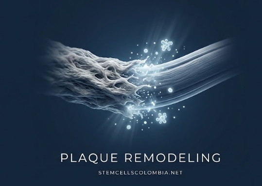
Healing Peyronie’s disease requires more than just masking the symptoms or surgically cutting out the problem. True structural repair involves active plaque remodeling. The goal is to reverse the fibrotic process at the cellular level and restore the natural elasticity of the tissues. This biological process relies on two primary mechanisms working together.
Inhibition of the Pro-Fibrotic Pathway
The first step in remodeling a plaque involves the targeted inhibition of the pro-fibrotic TGF-beta1 signaling pathway. By arresting the signals that tell the tissue to keep producing scar material, the cellular environment can begin to stabilize. Downregulating this pathway prevents further plaque propagation. More importantly, shutting off the TGF-beta1 signal encourages the overly active myofibroblasts to undergo apoptosis. Apoptosis is the programmed death of a cell. When these rogue, scar-producing cells naturally die off, the aggressive contraction of the tissue finally stops.

Upregulation of Matrix Metalloproteinases
Once the aggressive scarring signal is halted, the second mechanism takes over. This involves the upregulation and activation of matrix metalloproteinases, commonly referred to as MMPs. MMPs are specialized enzymes naturally found in the body that are capable of breaking down dense interstitial collagen bundles.
Specifically, enzymes like MMP-1, MMP-2, MMP-8, and MMP-13 are activated to selectively degrade the hardened scar tissue without harming the surrounding healthy anatomical structures. This enzymatic degradation clears away the rigid collagen blocks, re-establishing a dynamic equilibrium between tissue synthesis and tissue clearance.
As the plaque dissolves, the body has a chance to rebuild correctly. The local cellular environment can reorganize new collagen fibrils into functional, parallel alignments rather than chaotic scar bundles. Simultaneously, the tissue begins to promote the synthesis of fresh elastin, gradually restoring the essential tensile flexibility of the tunica albuginea.
Regenerative Protocols and Clinical Delivery
The scientific understanding of plaque remodeling has opened the door to advanced regenerative protocols. Traditional surgical methods often involve cutting or grafting the plaque, which can lead to a loss of penile length and a high risk of permanent nerve damage. Modern regenerative approaches take a more sophisticated, biological route to address the root cause of the fibrosis.
Cellular and Paracrine Interventions
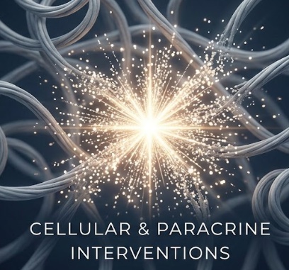
Cellular and paracrine interventions are currently at the forefront of this medical field. Mesenchymal stem cells, alongside their secretome and exosome derivatives, act as potent biological modulators. When introduced to the fibrotic environment, these biologic materials act as communication hubs. They release specific growth factors and anti-inflammatory cytokines that directly inhibit the stubborn TGF-beta1 pathway and actively stimulate local MMP production.
Precision Delivery Strategies
Delivering these biologics requires extreme precision. Simply injecting medication blindly into the tissue is not enough. Medical professionals utilize high-resolution, ultrasound-guided intralesional injections to place the biologic agents directly into the dense core of the fibrous plaque. This exact placement ensures that the active compounds are concentrated exactly where the structural tissue remodeling needs to occur.
Adjunctive Bio-Mechanical Modalities
Furthermore, leading regenerative clinics rarely rely on a single treatment approach. They combine these cellular therapies with adjunctive bio-mechanical modalities to maximize the results. Low-intensity extracorporeal shockwave therapy is frequently applied to the area to stimulate mechanotransduction. The sound waves help break up calcified deposits within the plaque and enhance local blood flow, which helps carry away the enzymatically degraded collagen.
Penile traction therapy is another critical addition to the protocol. Traction applies a gentle, sustained mechanical stress to the tissue. As the biologic treatments dissolve the plaque, the traction device guides the new collagen fibers into a straight, functional alignment, preventing the tissue from healing back into a curved shape.
Therapeutic Endpoints and Expected Outcomes
Patients undergoing regenerative plaque remodeling can track their progress through several specific, measurable therapeutic endpoints. Modern treatment focuses on clear clinical markers to ensure the therapy is working as intended.
Tracking Structural Metrics
The primary focus is always on structural metrics. Over the course of the treatment protocol, physicians expect to see a measurable reduction in the degree of penile curvature. High-resolution ultrasound imaging is used at various intervals to verify a physical decrease in overall plaque volume. Additionally, doctors look for a reduction in tunical thickening, confirming that the healthy tissue is returning to its normal anatomical state.

Symptom Resolution and Functional Recovery
Beyond structural changes, symptom resolution is a critical marker of success. For men treated in the early, active phase of Peyronie’s disease, alleviating the severe inflammatory pain experienced during erection and flaccidity is a major clinical milestone.
As the tissue regains its elasticity and the curvature diminishes, patients typically experience a significant functional recovery. This recovery is evaluated by noting improvements in penetration capability and by tracking scores on standard diagnostic questionnaires, such as the International Index of Erectile Function. Ultimately, the successful degradation of collagenous fibrotic plaques allows men to enjoy long-term tissue compliance. By addressing the exact biological causes of penile fibrosis, modern regenerative medicine provides a safe, highly effective pathway back to normal function.